When you arrive, please report to the reception desk on the ground floor of the Richard Desmond Children's Eye Centre (RDCEC).
What is visual electrophysiology?
When you look at something, an image of the object is projected onto the retina at the back of your eye. The retina converts this optical image into very small electrical signals, which pass along the optic nerve to the brain, where the sensation of ‘seeing’ occurs. Visual electrophysiology measures these very small signals produced by the eye and the brain.
Why is my child being referred?
A doctor has referred your child for electrophysiology tests to assess how their visual system manages visual information. The tests can help to diagnose the cause of a visual problem. They are also useful for monitoring the development of a visual disorder or the effects of any treatment. These tests are particularly useful as young infants and children are often unable to communicate or describe in detail any problems they might have with the way they see.
How do we do the tests?
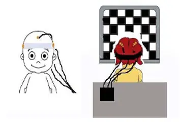
We record the small electrical signals from the eye and the brain in response to a flashing light or a pattern on a computer screen. These signals are recorded by small contacts that are placed on the surface of your child’s head and near the eye. The area under the contact will be gently cleaned with a gel before the contact is applied. This may be slightly uncomfortable. The recording of the activity of the retina is called the electroretinogram (ERG); the recording of the activity of the brain is called the visual evoked potential (VEP).
Visual evoked potential (VEP)
To record the VEP, small contacts are placed on your child’s head, using some paste. Your child will be asked to look at a light which will flash twice a second and at a moving black and white pattern on a TV screen.
At the end of the test, the contacts will be removed.
We try and remove all the paste from the hair with warm water but you will probably want to wash your child’s hair when you get home.
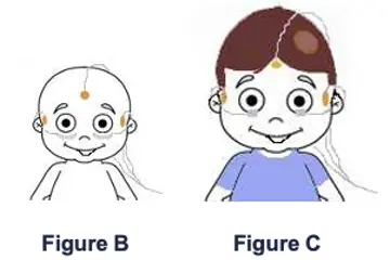
Electroretinogram (ERG)
To record the ERG in babies and young children, small contacts are placed at the side of the eye and under each eyelid (Figure B). The contacts at the side of the eye are kept in place with tape. In older children, thin thread-like contacts are placed over the lower eyelids (Figure C).
Your child will be asked to look at a light that flashes at different speeds and brightness levels. The contacts are then removed and the skin cleaned.
Will the tests hurt?
The tests do not hurt.
When recording an ERG in children over six years of age, we may use eye drops to dilate the pupils, similar to those used in the clinic, or very occasionally, anaesthetic (numbing) drops. The drops may cause a slight stinging sensation for a few seconds and your child’s vision may become blurred. Vision normally recovers within a couple of hours.
What will happen next?
The results of the tests will be analysed and a report will be sent to the consultant who referred your child for testing.
Author: Magella Neveu, Clinical Scientist-visual electrophysiology
Review date: October 2026