Check out the Ultrasound room in the Eyesite Virtual Eye Hospital
What is it?
You will know what an X-ray is. It’s a way of making a picture of the inside of your body to see, for instance, if you have a broken bone. Ultrasound is a bit similar. It is used to make a picture of the inside of your eye.
Just like an X-ray, ultrasound doesn’t hurt at all, takes only a few minutes, and you won’t need to be asleep while it happens.
How does it work?
Do you know how a bat can “see” in the dark by making high pitched sounds? The bat listens to the echoes, which tells it the position of nearby objects and food. Dolphins and whales can do this also.
Ultrasound works in a similar way. You close your eye and something that looks a bit like a small wand is moved over it. The wand sends ultrasound into your eye, but you won’t hear anything or feel any pain. The echoes bounce back to a computer. The computer uses the echoes to make images which show what the inside of your eye looks like.
Why do I have to have one?
Someone called an ophthalmologist may need to examine your eye. To do this, they will usually just shine a light into your eye. But if you have some blood in your eye, or a cataract, it can make it hard for them to get a really good look. So they will use ultrasound instead.
It might be necessary to know the length of your eye, from front to back. You couldn’t really use a tape measure to find out, could you? Again, ultrasound would be used.
What happens during the test?
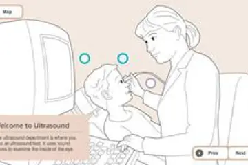
Examinations take just a few minutes to complete and are painless, with no anaesthesia required. Ideally the head should kept be as still as possible.
Children are examined whilst seated. Babies or young infants are generally examined whilst cradled on a parent’s lap but may be examined in a portable car seat or buggy.
A probe is smeared with a liquid gel and moved over the closed eyelid. The probe emits pulses of ultrasound into the eye and surrounding tissue. During the brief intervals of time between each pulse, the probe receives any echoes which scatter back towards it. The echoes are converted into electric signals which are displayed on screen as real-time images of the eye in cross section.
The operator performing the scan can make diagnoses from the moving images on the screen and digitally store any selected images.
Measurements of eye structures are generally made from any stored images once the examination is over. A report is produced immediately following the examination for patients to take back to the referring ophthalmologist.
What will happen after?
You will be sitting down during an ultrasound test. Afterwards you can see the images of the inside of your eye and the ophthalmologist will tell you and your parents what the pictures show.