From 4 March 2024, part of the pavement on Cayton Street will be closed.
This will impact where staff and patients cross between our trust and Private sites, and is due to external construction works taking place.
For patients
-

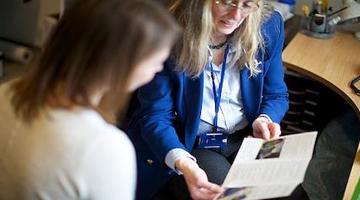
Plan your visit
Learn about everything you need to know about preparing for your appointment at Moorfields, including your arrival, parking and visitor information.
-

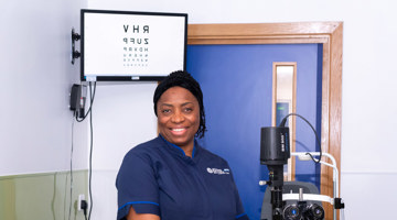
Our consultants
Learn more about our world-class and passionate consultants
-

Our locations
Find out more about our many Moorfields locations

Moorfields charities
-
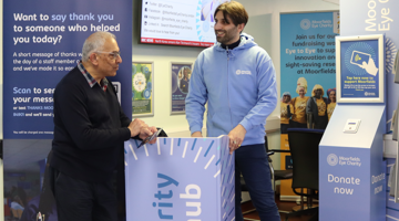
Moorfields Eye Charity
Moorfields Eye Charity is the leading charity in the UK funding research into eye health and innovation and improvement in patient care to help patients at Moorfields and globally.
-
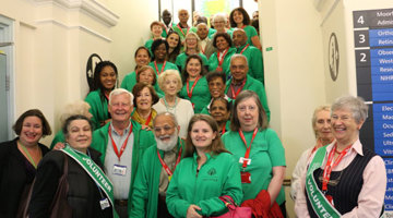
Friends of Moorfields
Our staff and volunteers provide a range of services to support the hospital, from guiding and directing patients to providing eye health information and reassuring patients during procedures.
Your support network
-
Eye clinic liaison officers (ECLOs)
Practical information and rehabilitation advice on living with sight loss.
-
Counsellors
Emotional support, information and advice for patients and their relatives from diagnosis to follow-up.
-

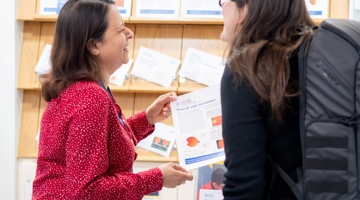
Patient advice and liaison service (PALS)
Providing confidential advice and support to resolve concerns about your care and guide you through our services.
About us

-
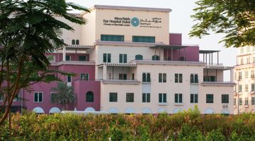
Moorfields UAE
Moorfields Eye Hospital Dubai is the first overseas branch of Moorfields. Moorfields Eye Hospital Centre Abu Dhabi, in partnership with United Eastern Medical Services, is the first Joint Venture of Moorfields.
-
Moorfields Private
Moorfields Private is the private division of Moorfields Eye Hospital.
The financial surplus from Moorfields Private is reinvested back into Moorfields Eye Hospital NHS Foundation Trust for the benefit of all patients.
-

Oriel - creating the centre for advancing eye health
Oriel is the joint partnership between Moorfields Eye Hospital NHS Foundation Trust, the UCL Institute of Ophthalmology and Moorfields Eye Charity that will move services from Islington to a new, integrated centre in Camden.
For Health Professionals
-

Single Point of Access
Single Point of Access (SPA) provides a first point of contact for people wishing to access adult community services across London and Hertfordshire.
-

Education and training
Moorfields Education works in partnership with UCL Institute of Ophthalmology to deliver world class continuing medical education.
-
Our consultants
Learn more about our consultants to refer patients to
More from Moorfields
-
Careers
Join our mission to be the leading international centre in the care and treatment of people with eye disorders, driven by excellence in research and education.